
Hiding in plain sight
A 50 yo female presents to the ED for abdominal pain. She has a history of multiple prior bowel surgeries, and a CT scan is requested. I do not see an acute finding on the CT, and as I am going through my search pattern, the pancreas looks normal.
But is the pancreas really normal?
On the lower keV images, there is an intensely enhancing mass at the undersurface of the pylorus/duodenal bulb. This is nicely depicted on the iodine map. On spectral curves, this is identical to the pancreas.
So yes, the pancreas is normal, but there is a small accessory pancreas adjacent to the pylorus/duodenal bulb. This was hiding in plain sight on multiple prior exams, and is nicely seen in retrospect on a previous MRI.
Here is a great paper on heterotopic pancreas. The stomach/duodenum are the most common site of this fairly common anomaly. Heterotopic pancreas can have the same complications as the normal pancreas including pancreatitis and neoplasia.
The heterotopic pancreas can be confused with all kinds of pathology. Spectral CT provides another way to confirm the diagnosis with a “tissue signature” in the form of spectral curves.
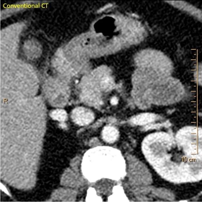
Conventional CT: Not much to talk about
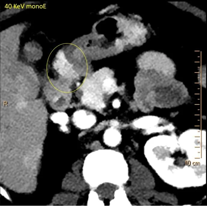
40 keV monoE: See the enhancing nodule at the undersurface of pylorus/duodenal bulb
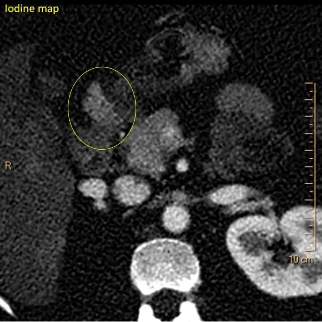
Iodine map shows uptake identical to the pancreas
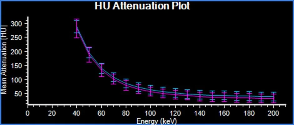
Spectral curves in the pancreas (blue) and the heterotopic pancreas (magenta) are identical!
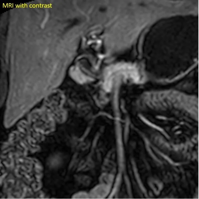
Previous MRI shows the heterotopic pancreas nicely. It was hiding in plain sight!