
Open your eyes, and you will see
76 yo F, past h/o lung cancer presents with flank pain and fever. Conventional CT obtained. Very subtle signs of pyelonephritis on the right, with cortical hypoperfusion.
Turn on spectral, and the cortical hypoperfusion is very easy to appreciate. But, lo and behold, a previously invisible gallstone is also now visible!
Spectral CT will make it easy to see what you need to see, but will also show you what you do not expect to see.
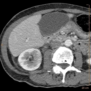
Conventional CT shows subtle hypoperfusion in the right kidney medially. Note the gallbladder looks normal.

Iodine map accentuates the renal hypoperfusion, consistent with pyelonephritis
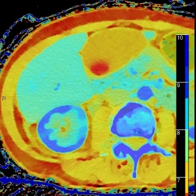
Overlay Z-effective image shows the renal cortical defect very well, but note a gallstone now pops into view!
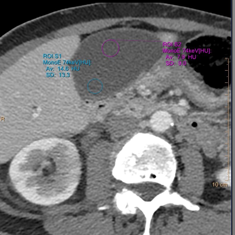
Attenuation of the gallstone and bile is nearly identical on conventional images
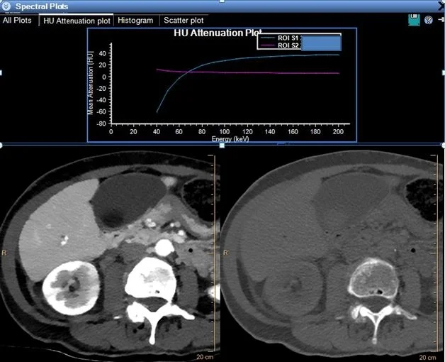
Behavior of gallstone and bile on spectral analysis is quite different. The curves intersect at about 70 keV, which is typical energy of conventional CT scan. Gallstones (blue curve) are strikingly hypodense on low energy relative to bile (magenta curve, also image on left) and high density on higher energy (image on right).