
A case of neck pain
65 yo F with neck pain for a few months. CT shows pathological fracture of C7 and T1, confirmed on MRI. A search for the primary ensued.
Conventional CT of the abdomen shows mild pancreatic ductal dilatation in the tail of the pancreas but is otherwise unremarkable. MRI shows a hypo enhancing mass in the pancreatic tail with restricted diffusion. Small (1.5 cm) pancreatic adenocarcinoma confirmed with endoscopic ultrasound and biopsy.
Review of 40 keV spectral images shows clear hypo-enhancing mass in pancreatic tail, corresponding excellently with mass seen on MRI. Spectral CT can be a life-saver in this situation.

Cervical spine CT shows pathological fracture of C7 and T1
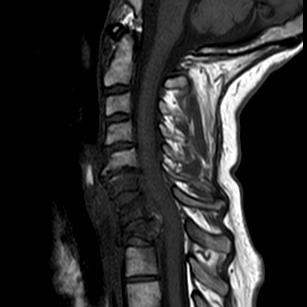
MRI confirms pathological fractures

Conventional CT shows very subtle lesion in pancreas, not prospectively call-able

40 keV mono-energy image shows clear lesion in pancreatic tail with upstream ductal dilatation
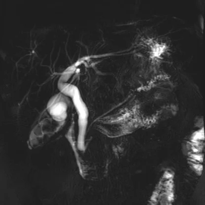
MRCP with focal ductal dilatation in the pancreatic tail shown very nicely
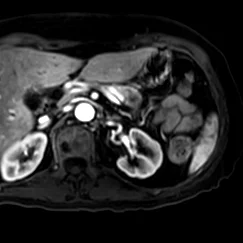
Contrast enhanced MR shows hypoenhancing pancreatic lesion

Diffusion wieghted MR (b=400) shows resricted difffusion in lesion

Mass biopsied on endocspoic ultrasound