
GI bleed
43 yo male with history of lymphoma and poor medical compliance presented with shortness of breath and dark stools. Hemoglobin came back at 3.0 mg/dL.
Conventional CT shows complex fistula in LUQ involving spleen, stomach and colon. On spctral analysis, virtual non-contrast image shows increased contrast between myocardium and blood pool, consistent with anemia.
Ill-defined density in the posterior aspect of the fistula shows clear iodine uptake and is consistent with active GI bleed. Patient was stabilized and taken to the operating room, with extensive surgery confirming B-cell lymphoma.
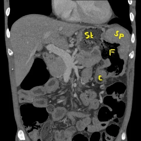
Conventional CT (coronal image) shows fistula (F) between the stomach (St), spleen (Sp) and colon (C)

Virtual non-contrast image shows increased contrast between LV myocardium (48 HU) and blood pool (27 HU), consistent with anemia
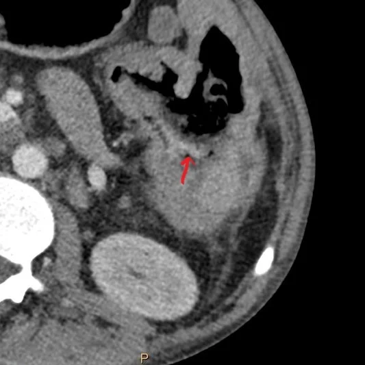
Conventional CT (axial plane) shows ill-defined density (red arrow) in the posterior aspect of fistula
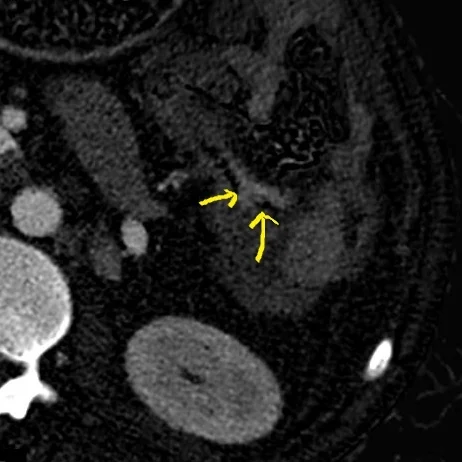
Iodine uptake (yellow arrows) confirms GI bleed