
The case of the wandering woman
A young female was found behaving erratically, having left her vehicle after a crash. A head CT and a CT scan of the chest, abdomen and pelvis obtained for trauma.
Surprisingly, the head CT showed a left middle cerebral artery stroke, with a dense MCA sign and loss of cortico-medullary differentiation. The trauma CT showed subtle bilateral pulmonary emboli. No other acute finding.
Further workup showed a patent foramen ovale. The working hypothesis is she had pulmonary emboli, and then the stroke secondary to right to left shunt through the PFO. Interetestingly there is no clear contralateral weakness on exam.
This case shows the amazing ways in which disease processes can manifest, and quite often, all is not as simple as it seems.
Of course, you can also appreciate the power of spectral CT in diagnosing subtle pulmonary emboli: vastly improving detection and the diagnostic confidence of the radiologist.
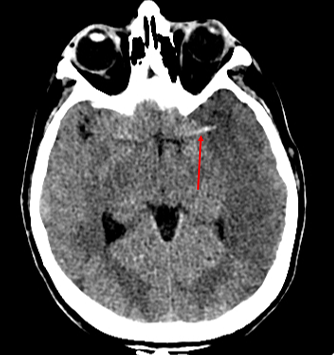
Non-contrast head CT shows large left MCA territory infarct. Note the dense MCA sign.
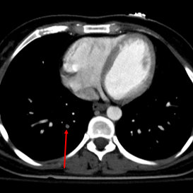
Trauma CT shows subtle right lower lobe PE. This should be easy to pick up on conventional CT.

This lingular PE should be relatively easy too..
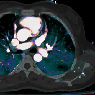
....notice how well the perfusion defect on iodine overlay stands out in the corresponding vascular territory

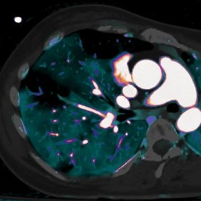
There is a PE in the left lower lobe lateral segment, but no matter how good a day you are having, you are not going to see it

Impossible to miss it on iodine overlay!

Another tiny PE in the right lower lobe on an oblique axial image...
And the iodine overlay again makes it impossible to miss!