
The hot appendix: part 2
48 year old female with chronic intermittent abdominal pain, presents to the ED with acute onset right lower quadrant pain. Peritoneal signs on exam, and white count elevated to 13,000. No fever.
Conventional CT shows peritonitis, with diffuse peritoneal thickening, omental stranding and small free fluid. the appendix is thick and inflamed, but is it perforated? The lining does not show a discontinuity.
Fortunately, spectral CT comes to the rescue! Obvious defect in appendiceal wall iodine uptake, consistent with perforation.
Patient taken to the OR, where perforated appendicitis with peritoneal purulence was confirmed. Iodine maps can significantly enhance detection of perforations in the GI tract, including the appendix.

Conventional coronal CT with peritoneal thickening (red arrows), consistent with peritonitis
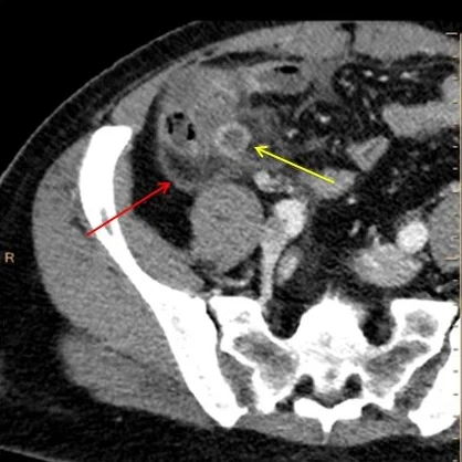
Axial conventional CT shows dilated appendix (yellow arrow). Adjacent fluid and inflammation, with red arrow showing the thick peritoneal lining.
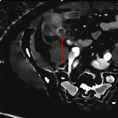
Iodine density image. Note focal discontinuity in appendix enhancement (red arrow). This is consistent with perforation.
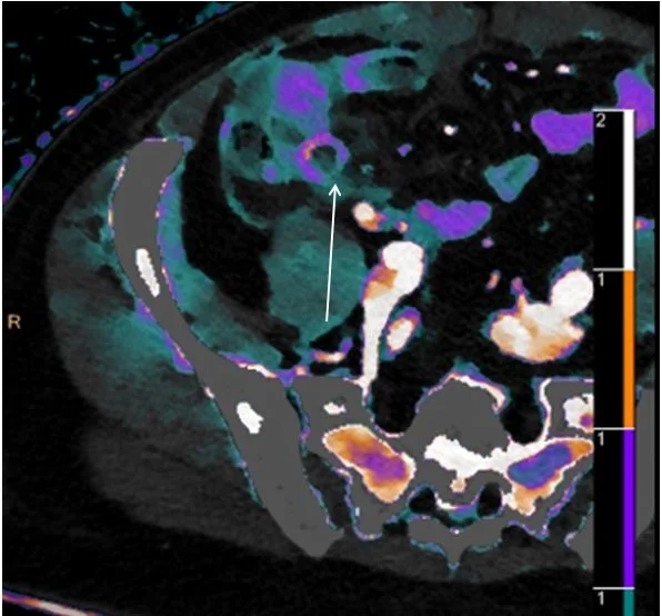
Iodine density overlay. Note focal discontinuity in appendix enhancement (white arrow).