
A case of a lung infiltrates
A young female immigrant presented with productive cough and fever. She had a history of previous peritoneal TB, and AIDS, and pulmonary TB was suspected with diffuse infiltrates seen on chest X-ray. CT scan of the chest performed with IV contrast showed diffuse infiltrates concerning for pneumonia.
Closer look at some of the non-confluent upper lobe infiltrates show a "flame-shaped appearance". On spectral analysis, these nodules show substantially higher iodine uptake than background lung.
Subsequent bronchoscopy showed reddish and purplish plaques in the airway, consistent with Kaposi's sarcoma.
Intense iodine uptake in lung lesions is compatible with the highly vascular nature of Kaposi sarcoma lesions. TB is very hypovascular in my experiece (as are most infections).
Unfortunately, patient passed away from complications.
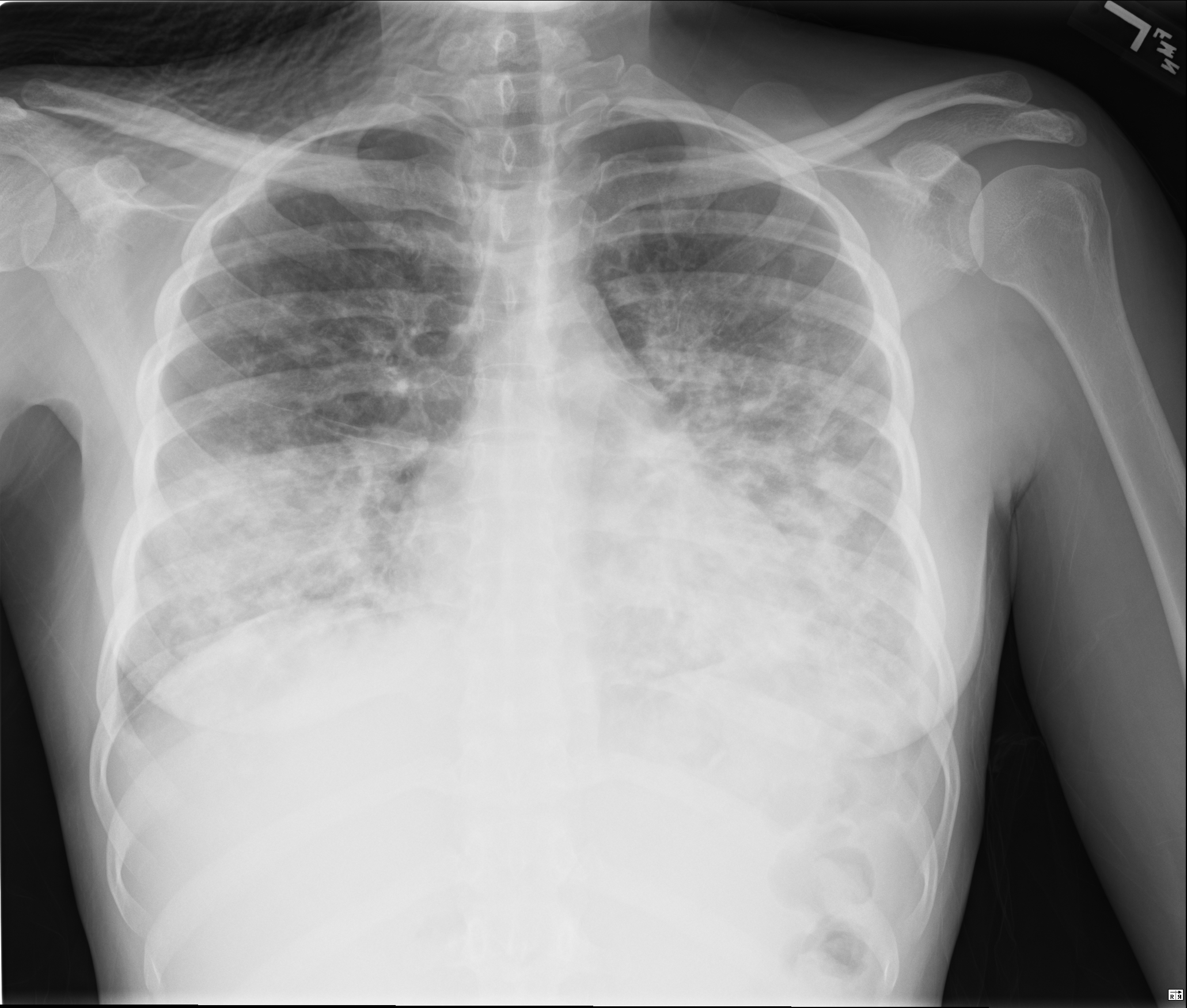
CXR with diffuse infiltrates

Conventional CT (lung windows)
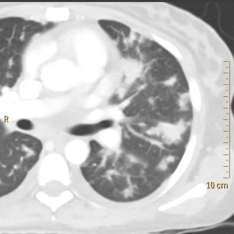
Infiltrates in left lung have "flame shaped" appearance
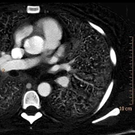
INcreased iodine uptake in the nodules

Iodine density is more than double of normal lung (2 mg/mL in lesion vs 1 mg/mL in normal lung)
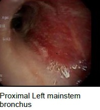

Bronchoscopy shows plaques of Kaposi sarcoma