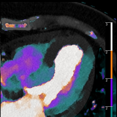
Myocardial infarction
45 yo M with acute chest pain. CT chest done to rule out dissection.
Conventional CT image through left ventricle looks within normal limits.
Spectral images show large perfusion defect in the distal antero-septal wall. Note iodine uptake is essentially zero (about 1.15 mg/mL in normal myocardium).
Patient had a LAD infarct with stent placement done 3 days prior to CT.


