
Tumor thrombus
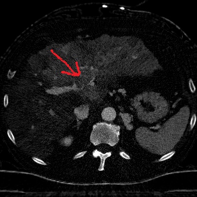
Routine CT shows heterogenous left lobe of liver, with filling defect in left portal vein.
Iodine uptake in thrombus confirms tumor, there is also diffuse HCC in left lobe.
Virtual non-contrast also shows large blood clot adjacent to left lobe, likely from a bleeding HCC.

Conventional CT with filling defect in portal vein (yellow arrow)
Iodine map shows perfusion in portal vein clot, consistent with tumor thrombus
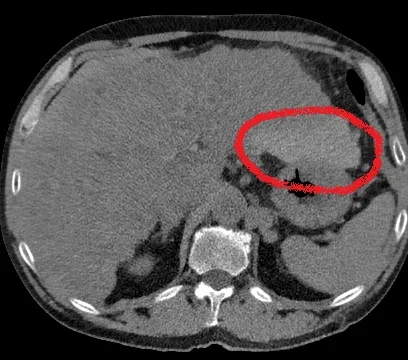
Virtual non-contrast shows bleed adjacent to the left lobe