
Why spectral CT?
72 year old male, with chronic hepatitis C, genotype 6 (could not complete treatment), presents with weight loss. Past history of HCC with chemoebolization. AFP is normal. A CT scan with liver protocol is ordered. Turns out he also has chronic renal failure with eGFR in the 30’s. BMI is 21.
Patient is in the department for the scan. We decide to use Spectral CT to reduce dose of contrast. After a true non-contrast scan (done as he had previous chemoembo), the multiphasic scan is done with 35 mL of omnipaque 350.
On images below, it is obvious Spectral CT helps in many ways. On the non-contrast phase, the 2.4 cm lesion in segment 6 is so much more obvious on the electron-density image (you know I like EDW). On the arterial phase and delayed phase, the lesion shows enhancement and washout respectively, and both are so much better seen on the 40 keV monoenergy reconstruction. This is a liRADS 5 lesion(definite HCC).
But wait, there is more. There is a subtle additional focus on arterial enhancement in segment 4A. This simply cannot be seen on conventional images, but cannot be missed on 40 keV image and iodine map. On the delayed scan, this lesion shows up much better on the electron density map.
The total DLP was about 660 mGy*cm, which is not bad for a multiphasic scan. So we did not zap the patient in the attempt to reduce contrast dose!
This case defines the utility of Spectral CT to me: being able to adjust dose of contrast to the lowest possible, and using the tools provided by spectral CT to give the patient and provider an accurate diagnosis the first time, and every time.
So what is your reason to not use Spectral CT?
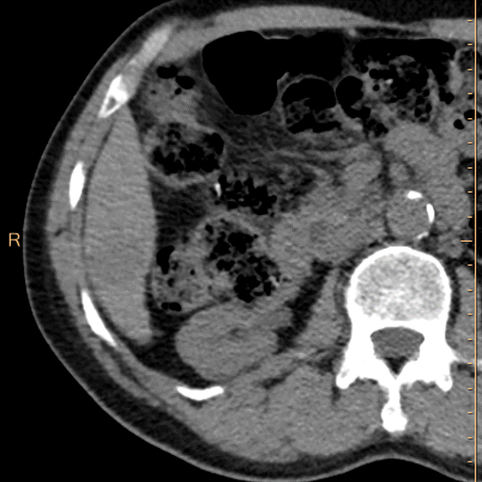
Non-contrast CT. Can you see the lesion?
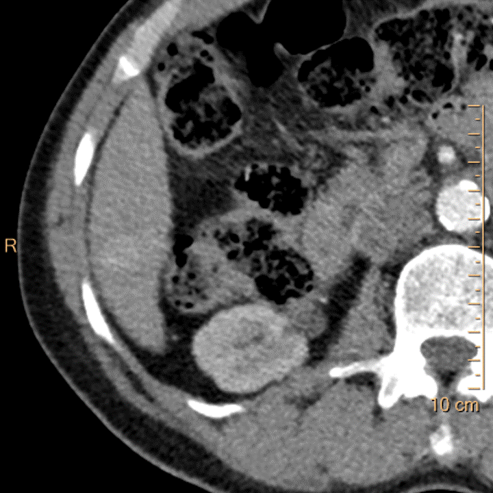
Electron density: Can anyone miss the lesion?
Electron density overlay: Just showing off now.
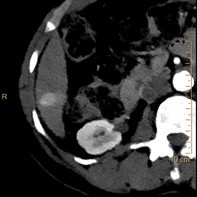
Arterial phase, conventional image. Scan done with 35 mL contrast. See the enhancement?
Arterial phase, 40 keV mono-E image. No comment needed.

Arterial phase, iodine map.
Portal venous phase, conventional phase. You cannot assess for washout, which is one of the major criteria for LiRADS.
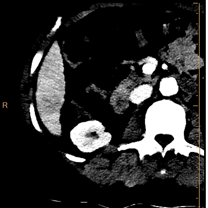
Portal venous phase, 40 keV mono-E. The washout is obvious!

PVP, electron density weighted image. See the lesion very well. Not sure this represents washout, as EDW seems to reflect non-contrast image quite well.
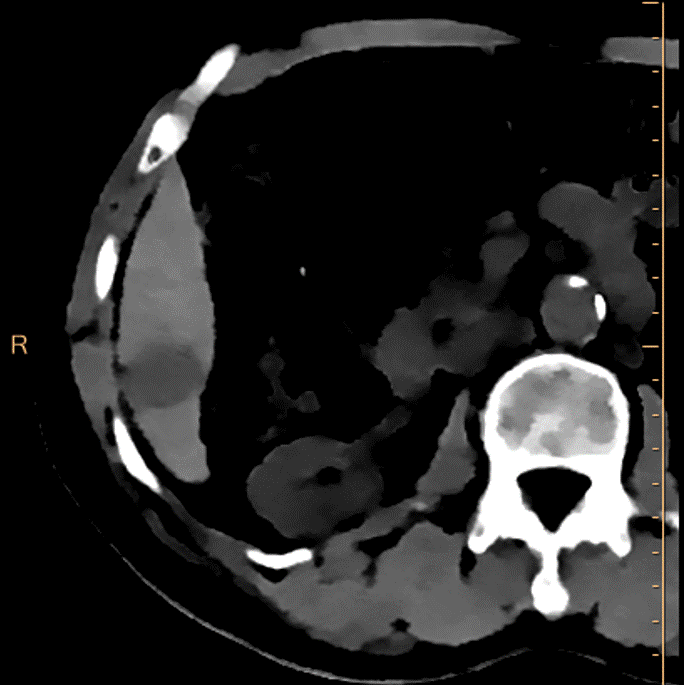
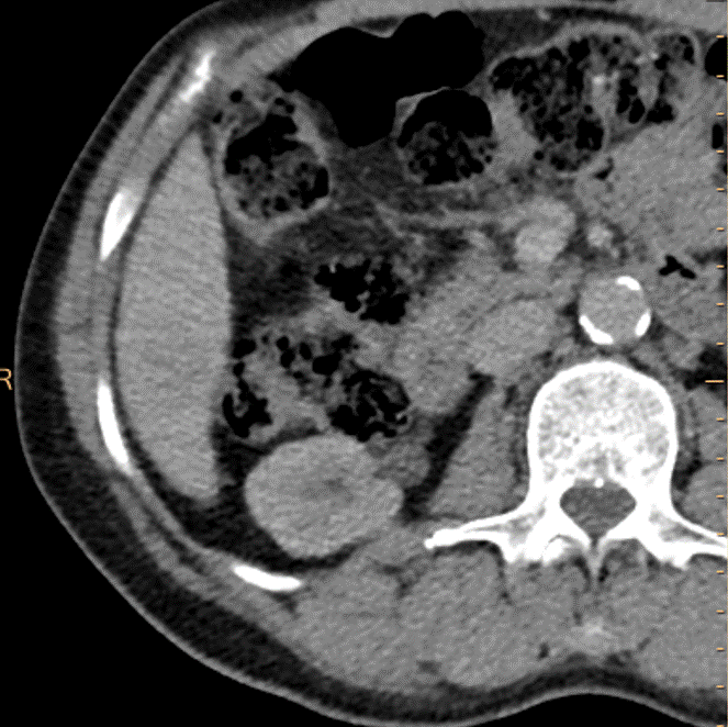
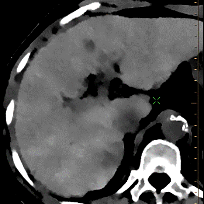
Conventional CT, arterial phase, higher up in the liver. This is unremarkable.
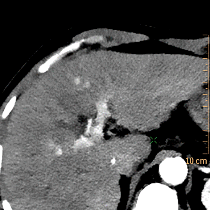
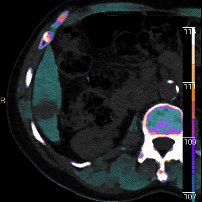
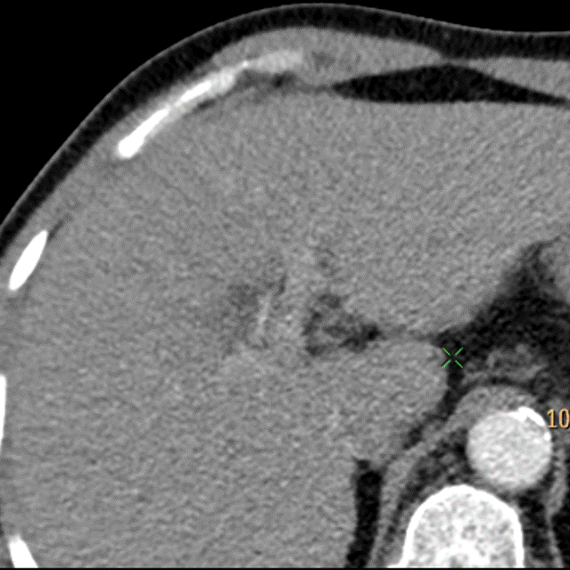
Arterial phase, 40 keV image. See the 2 dots of enhancement?
Delayed phase, electron density: The lesion shows up very well. Again, not sure this qualifies as true washout. In any case there is no capsule. I gave this a LiRADS 4, suspicious for HCC.