
Renal lesions made easy!
58 year old male, with a history of >30 prior abdominal surgeries presents with abdominal pain. A routine abdominal CT is obtained which shows a small bowel obstruction. Also noted is a 2.4 cm right kidney lesion.
Most abdominal CT scans in the ED are done in a single portal venous phase, and it can be difficult to definitively characterize renal lesions. No longer! Spectral CT can make renal lesions really easy!
In this case, there is obvious iodine uptake in the kidney lesion, and the lesion shows >90 HU enhancement relative to the virtual non-contrast. All this can be done on PACS itself, without needing any fancy processing. The PACS screenshot below illustrates the point.
MRI confirmed the findings, but was likely superfluous. Patient will follow up to discuss options.
Spectral CT can help us get it right, the first time, and every time!

Conventional CT: Subtle right kidney lesion. Note dilated loops of small bowel.
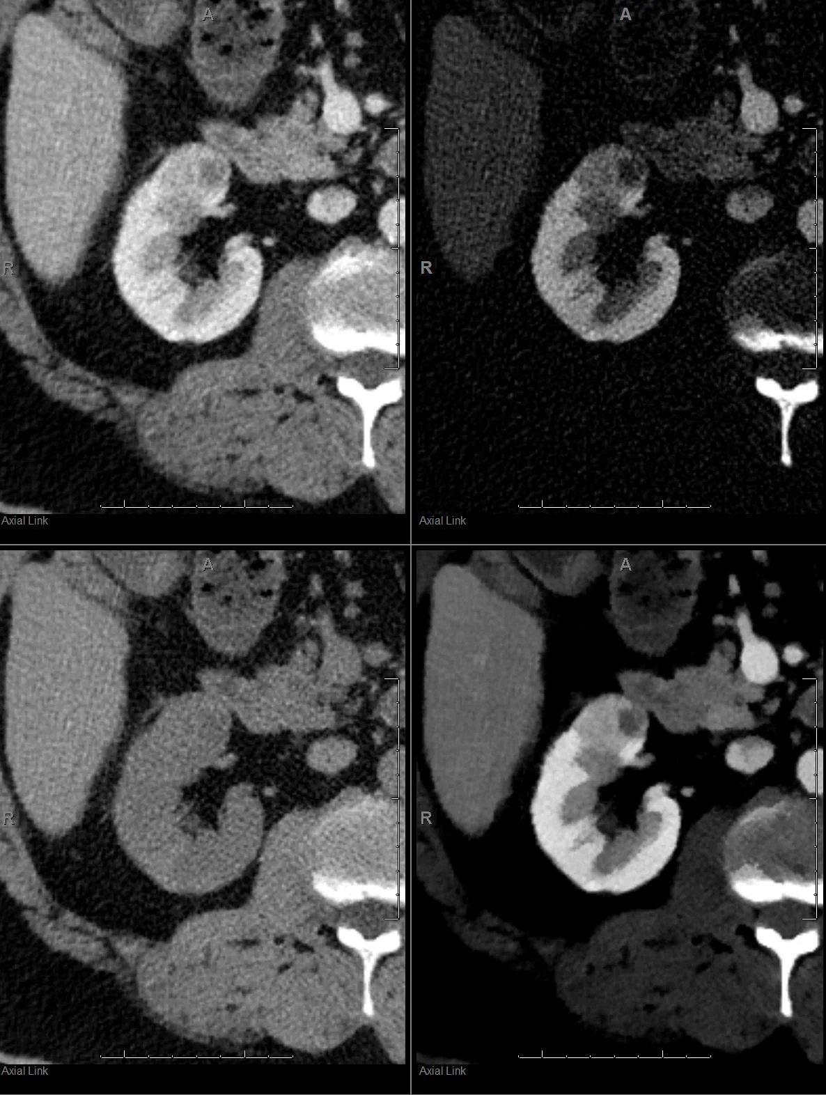
Screenshot from PACS (clockwise from top left): Conventional CT, iodine map, 40 keV mono-energy image and virtual non-contrast. The lesion is a renal cell carcinoma, and all you need is one glance at the spectral recons! At Hennepin, all these recons are automatically generated by scanner and exported to PACS.

More sophisticated measurements like iodine uptake can be done on dedicated thin-client workstation. Note that the iodine uptake in this lesion in 4.12 mg/mL, which is quite high, and suggests a clear cell RCC

MRI with contrast confirms what we already know. Notice how similar MRI looks to the iodine map.

Another PACS screenshot (different patient, clockwise from top left): Conventional CT with 82 HU indeterminate left kidney lesion, 40 keV mono-E image, iodine map with no iodine uptake at all, and virtual non-contrast with attenuation of 77 HU. This is a hemorrhagic cyst! You just saved the system an MR scan, and more importantly, saved this human being the anxiety and pain of an additional exam.