
Liver abscess
50 year old male presented with right upper quadrant pain. On exam patient was febrile, and lab evaluation showed elevated LFT's and leukocytosis.
Ultrasound showed a large hypo-echoic liver lesion. CT scan showed a large ill-defined liver lesion, considered to be abscess versus necrotic neoplasm. MRI was then performed with findings compatible with an abscess. 40 mL purulent fluid was removed under ultrasound guidance and drain placed.
On spectral analysis, the liver lesion shows complete absence of iodine uptake except for a shaggy rim. This is well seen on 40 keV image, with vastly superior contrast resolution, and replicates findings on MRI.
Careful analysis of spectral images may preclude need for additional MR imaging, thus adding value.

Ultrasound shows large hypoechoic liver lesion
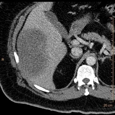
COnventional CT shows large ill-defined liver mass

Iodine map shows liver lesion is a "black hole" except for a shaggy margin which shows low iodine uptake, this appearance is very suggestive of an abscess

40 keV monoenergy image shows internal character of abscess very well
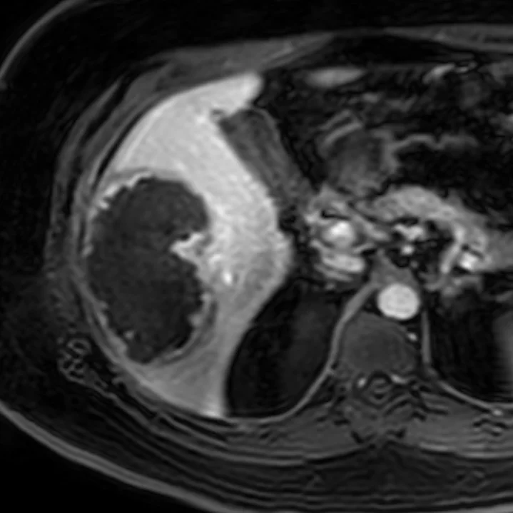
Post-contrast MRI reflects the findings on iodine map